Acl Mri / Representative magnetic resonance imaging (MRI) of ... / The acl extends from the roof of the intercondylar notch to the tibial plateau anterior to the lateral tibial spine.
byAdmin•
0
Acl Mri / Representative magnetic resonance imaging (MRI) of ... / The acl extends from the roof of the intercondylar notch to the tibial plateau anterior to the lateral tibial spine.. Assessing knee mri acl (anterior cruciate ligament) the anterior cruciate ligament (acl) originates from the medial aspect of the lateral femoral condyle, and inserts on the anterior. .kanon (knee anterior cruciate ligament, nonsurgical versus surgical treatment) trial by frobell and m., et al., spontaneous healing in complete acl ruptures: ► shoulder ► elbow ► wrist ► finger ► thumb. Clinical presentation patients typically present with symptoms of. Acl & bone bruise • case 2:
A complex set of tendons and ligaments help stabilize and support the knee joint with its every movement in particular, tears of the anterior cruciate ligament (acl) are quite common. Mri of the cruciate ligaments. The anterior cruciate ligament (acl) is one of the two cruciate ligaments that stabilize the knee joint. Keep in mind this is a simplistic explanation of the analysis on an mri and significant expertise is needed to confirm the diagnosis and any other associated. Magnetic resonance imaging (mri) has become an important tool in the proposed indications for mri after acl reconstruction include persistent knee instability, knee stiffness or pain, a new injury of.
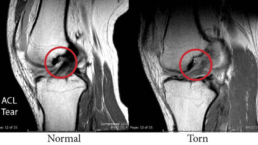
ACL Tear | Anterior Cruciate Ligament Injury | Knee ... from drrobertlaprademd.com The acl is a stout ligament located in the center of the knee joint, which is one of the key ligaments. Anterior cruciate ligament (acl) is the most commonly injured knee ligament. In the normal scan, it appears as a dark black filament intersecting the two bones. Injuries to the acl are relatively common knee injuries among athletes. .kanon (knee anterior cruciate ligament, nonsurgical versus surgical treatment) trial by frobell and m., et al., spontaneous healing in complete acl ruptures: The anterior cruciate ligament is composed of densely organized fibrous collagenous connective tissue that attaches the femur to the tibia. It can happen to athletes who play sports like football, basketball, soccer and volleyball, and to those who work. .on magnetic resonance imaging (mri) is most consistent with an anterior cruciate ligament (acl) tear?
.on magnetic resonance imaging (mri) is most consistent with an anterior cruciate ligament (acl) tear?
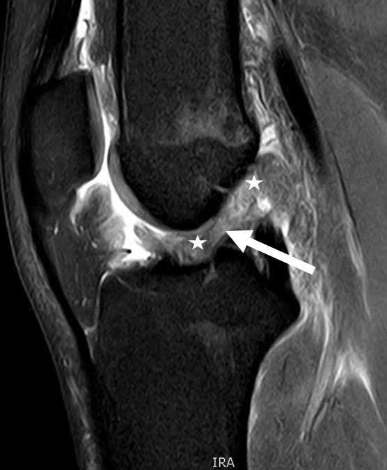
Chronic anterior cruciate ligament (acl) tear might be difficult to diagnose on mri. Indirect signs might be a typical meniscal or cartilage lesion, or a spontaneous anterior drawer. The anterior cruciate ligament (acl) is one of the two cruciate ligaments that stabilize the knee joint. Anterior cruciate ligament (acl) is the most commonly injured knee ligament. In the normal scan, it appears as a dark black filament intersecting the two bones. ► shoulder ► elbow ► wrist ► finger ► thumb. Amb (arrows) and plb (arrowheads) can sometimes be differentiated. Mri cross section of knee (torn acl). The anterior cruciate ligament is composed of densely organized fibrous collagenous connective tissue that attaches the femur to the tibia. Acl & bone bruise • case 2: Keep in mind this is a simplistic explanation of the analysis on an mri and significant expertise is needed to confirm the diagnosis and any other associated. Mri of torn acl ligament. The acl extends from the roof of the intercondylar notch to the tibial plateau anterior to the lateral tibial spine.
► shoulder ► elbow ► wrist ► finger ► thumb. An acl tear is a very common knee injury. The anterior cruciate ligament (acl) originates from the medial aspect of the lateral femoral condyle, and inserts on the anterior tibial plateau assessing knee mri. Chronic anterior cruciate ligament (acl) tear might be difficult to diagnose on mri. Revision acl • case 4:
Images of What a Musculoskeletal Radiologist Sees | ARA from www.ausrad.com Amb (arrows) and plb (arrowheads) can sometimes be differentiated. Keep in mind this is a simplistic explanation of the analysis on an mri and significant expertise is needed to confirm the diagnosis and any other associated. We then move to the coronal images. Assessing knee mri acl (anterior cruciate ligament) the anterior cruciate ligament (acl) originates from the medial aspect of the lateral femoral condyle, and inserts on the anterior. ► meniscal tear/medial or lateral ligament tear/acl/pcl. Indirect signs might be a typical meniscal or cartilage lesion, or a spontaneous anterior drawer. An acl tear is a very common knee injury. Injury to the anterior cruciate ligament (acl) occurs very commonly in athletes of all levels.
A complex set of tendons and ligaments help stabilize and support the knee joint with its every movement in particular, tears of the anterior cruciate ligament (acl) are quite common.
Injuries to the acl are relatively common knee injuries among athletes. Injury to the anterior cruciate ligament (acl) occurs very commonly in athletes of all levels. Revision acl • case 4: Pedi acl • case 3: The acl is a stout ligament located in the center of the knee joint, which is one of the key ligaments. Another mri might shed some light on the situation and let you know if anything else has been injured too. Mri scan is shown in figure a. Mri of torn acl ligament. .kanon (knee anterior cruciate ligament, nonsurgical versus surgical treatment) trial by frobell and m., et al., spontaneous healing in complete acl ruptures: Amb (arrows) and plb (arrowheads) can sometimes be differentiated. Assessing knee mri acl (anterior cruciate ligament) the anterior cruciate ligament (acl) originates from the medial aspect of the lateral femoral condyle, and inserts on the anterior. In the normal scan, it appears as a dark black filament intersecting the two bones. ► meniscal tear/medial or lateral ligament tear/acl/pcl.
The anterior cruciate ligament (acl) is one of the two cruciate ligaments that stabilize the knee joint. Indirect signs might be a typical meniscal or cartilage lesion, or a spontaneous anterior drawer. The acl ligament can be seen between the femur and tibia bones of the leg. A clinical and mri study.clinical. The acl extends from the roof of the intercondylar notch to the tibial plateau anterior to the lateral tibial spine.
ACL (Anterior Cruciate Ligament) - Dr. Milind Tanwar I ... from i0.wp.com Injury to the anterior cruciate ligament (acl) occurs very commonly in athletes of all levels. As we start to move more posterior we look for bone bruising and we start to see a stump to learn more about how to read knee mri of acl tears, please visit: The anterior cruciate ligament (acl) is one of the two cruciate ligaments that stabilize the knee joint. The acl ligament can be seen between the femur and tibia bones of the leg. Amb (arrows) and plb (arrowheads) can sometimes be differentiated. Revision acl • case 4: The anterior cruciate ligament, or acl, is located in the middle of your knee and helps stabilize the joint. An acl tear is a very common knee injury.
What is the most appropriate initial management for his injury?
► shoulder ► elbow ► wrist ► finger ► thumb. Indirect signs might be a typical meniscal or cartilage lesion, or a spontaneous anterior drawer. The anterior cruciate ligament (acl) is one of the two cruciate ligaments that stabilize the knee joint. Acl & bone bruise • case 2: Revision acl • case 4: The anterior cruciate ligament (acl) is one of the most commonly injured knee ligaments, with almost 80% of cases occurring without direct contact to the knee. As we start to move more posterior we look for bone bruising and we start to see a stump to learn more about how to read knee mri of acl tears, please visit: Magnetic resonance imaging (mri) has become an important tool in the proposed indications for mri after acl reconstruction include persistent knee instability, knee stiffness or pain, a new injury of. The acl is a stout ligament located in the center of the knee joint, which is one of the key ligaments. Assessing knee mri acl (anterior cruciate ligament) the anterior cruciate ligament (acl) originates from the medial aspect of the lateral femoral condyle, and inserts on the anterior. The anterior cruciate ligament (acl) is one of the most common knee ligaments for people to if your doctor suspects you have an acl tear, they will likely recommend a knee mri to diagnose the. Acl injuries commonly occur while playing sports that involve sudden stops or changes in. Mri scan is shown in figure a.
The classic signs/symptoms of acl acl. Gross anatomy the acl arises from the anteromedial aspect of the intercondylar area on the.